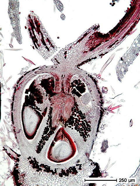
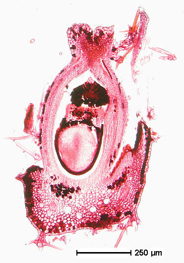
Pistillate Flower - Internal Anatomy
click on the small images to open larger format images
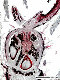
 Sections by Jenna L. Froelich (left, oblique longitudinal
section) and Michael A. Terry (right, longitudinal section)
Sections by Jenna L. Froelich (left, oblique longitudinal
section) and Michael A. Terry (right, longitudinal section)
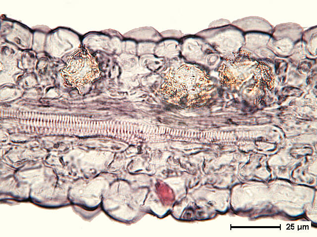
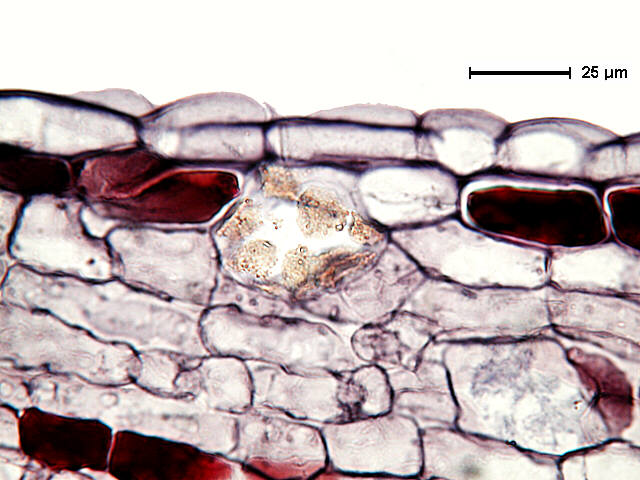
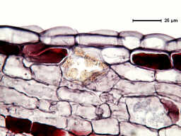
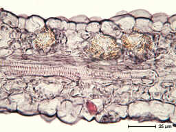 Section by Marnie J. Freeman, transverse section of
distal region of sepal; secretory idioblasts with amber-colored
granular deposits are charasteric of distal regions of the pistillate
flower sepals.
Section by Marnie J. Freeman, transverse section of
distal region of sepal; secretory idioblasts with amber-colored
granular deposits are charasteric of distal regions of the pistillate
flower sepals.
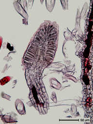
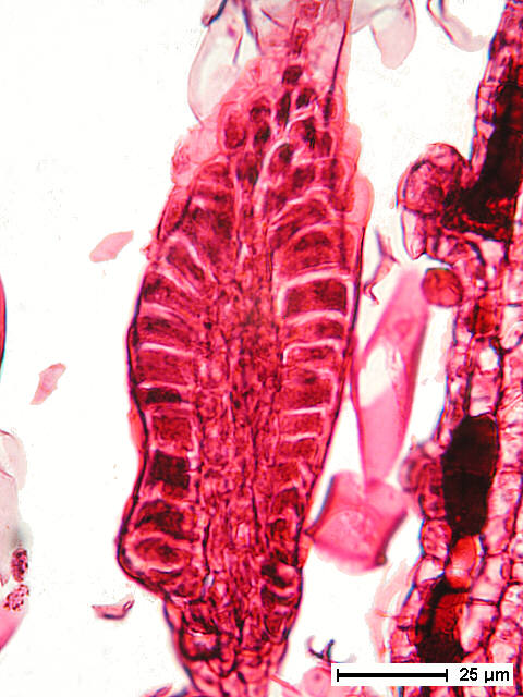
 Sections by Jenna L. Froelich (left) and Michael A.
Terry (right); longitudinal sections of petals; the palisade-like
epidermis resembles that of basilaminar glands and glandular foliar
teeth, suggesting a secretory function for the narrow, spike-like
petals.
Sections by Jenna L. Froelich (left) and Michael A.
Terry (right); longitudinal sections of petals; the palisade-like
epidermis resembles that of basilaminar glands and glandular foliar
teeth, suggesting a secretory function for the narrow, spike-like
petals.
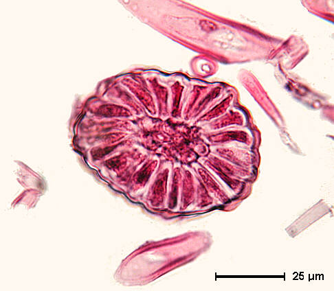
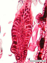
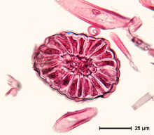 Section by Marnie J. Freeman; transverse section of
petal; the palisade-like epidermis resembles that of basilaminar
glands and glandular foliar teeth, suggesting a secretory function
for the narrow, spike-like petals.
Section by Marnie J. Freeman; transverse section of
petal; the palisade-like epidermis resembles that of basilaminar
glands and glandular foliar teeth, suggesting a secretory function
for the narrow, spike-like petals.
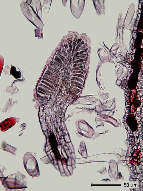
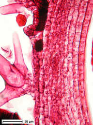
 Section by Jenna L. Froelich; style base; note secretory
idioblast with amber-colored granular deposits.
Section by Jenna L. Froelich; style base; note secretory
idioblast with amber-colored granular deposits.

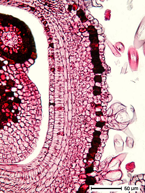
 Sections by Marnie J. Freeman (left) and Michael A.
Terry (right); ovary wall; note stellate trichomes on external
surfaces; the innermost two layers of the ovary wall will differentiate
into sclereids instrumental in the explosive dehiscence of the
mature fruits.
Sections by Marnie J. Freeman (left) and Michael A.
Terry (right); ovary wall; note stellate trichomes on external
surfaces; the innermost two layers of the ovary wall will differentiate
into sclereids instrumental in the explosive dehiscence of the
mature fruits.