Lamina - Internal Anatomy
click on the small images to open larger format images
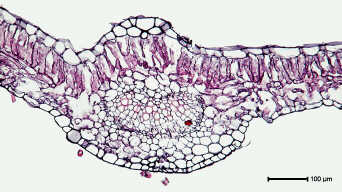
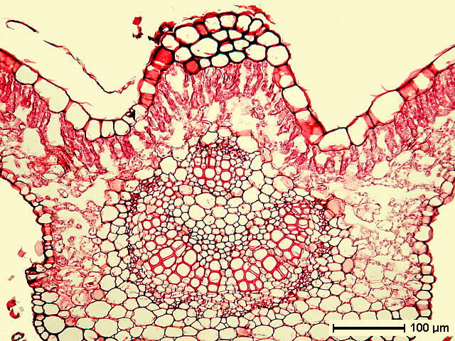
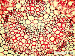
 Primary vein. Sections by Ali G. Davalos (left) and
Jeremiah E. McNamara (right). Note continuity of mesophyll over
the upper (adaxial) surface of the primary vein. Vascular tissue
of smaller leaves or distal regions of large leaves consist of
simple arcs of xylem and phloem (left); large leaves and their
basal regions often have an additional small bundle located above
the large arc.
Primary vein. Sections by Ali G. Davalos (left) and
Jeremiah E. McNamara (right). Note continuity of mesophyll over
the upper (adaxial) surface of the primary vein. Vascular tissue
of smaller leaves or distal regions of large leaves consist of
simple arcs of xylem and phloem (left); large leaves and their
basal regions often have an additional small bundle located above
the large arc.
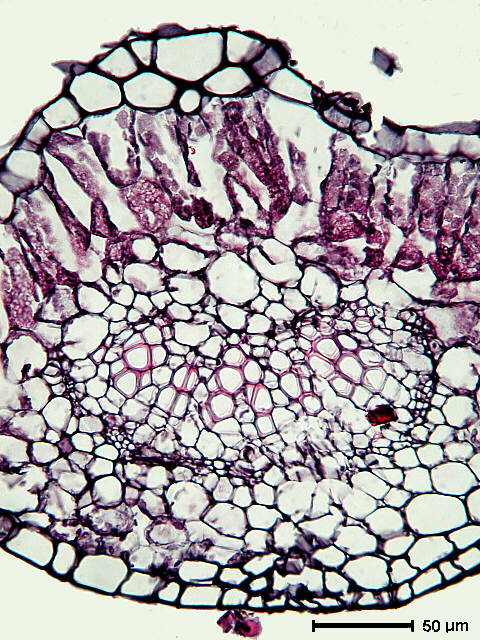
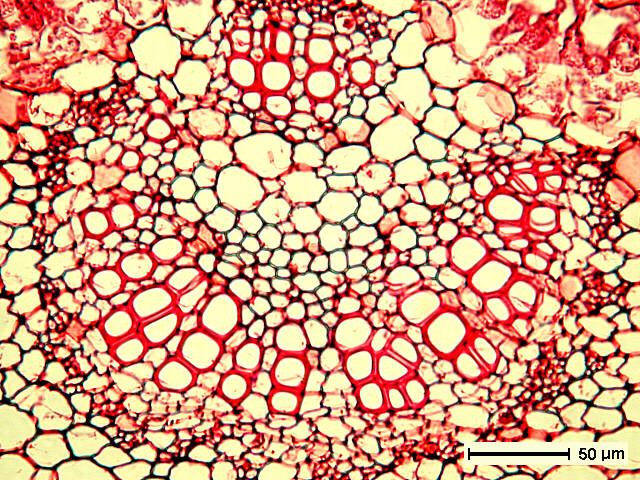
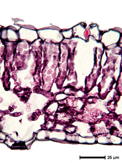
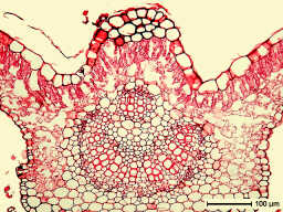
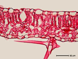
 Sections by Ali G. Davalos (left) and Jeremiah E. McNamara
(right); detials of primary vein.
Sections by Ali G. Davalos (left) and Jeremiah E. McNamara
(right); detials of primary vein.
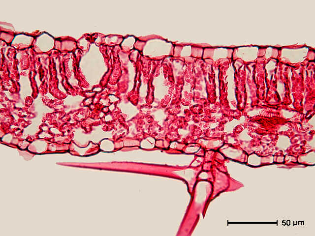
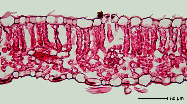
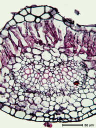
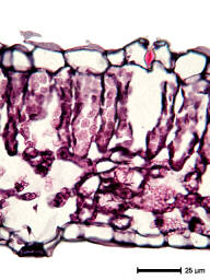
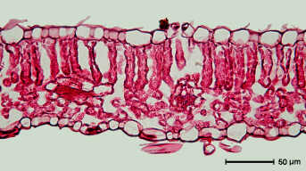 Sections by Jeremiah E. McNamara (left); note uniseriate
epidermides, amphistomatic lamina, and single palisade layer.
Also note absence of druse crystals; alum mordant used in the
hematoxylin-safranin combination characteristically dissolves
calcium oxalate crystals.
Sections by Jeremiah E. McNamara (left); note uniseriate
epidermides, amphistomatic lamina, and single palisade layer.
Also note absence of druse crystals; alum mordant used in the
hematoxylin-safranin combination characteristically dissolves
calcium oxalate crystals.
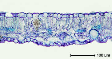
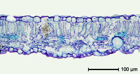
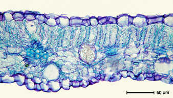 Sections by Susan B. Davidge (right); note uniseriate
epidermides, amphistomatic lamina, and single palisade layer.
Toluidine blue O stain technique leaves druse crystals intact.
Sections by Susan B. Davidge (right); note uniseriate
epidermides, amphistomatic lamina, and single palisade layer.
Toluidine blue O stain technique leaves druse crystals intact.
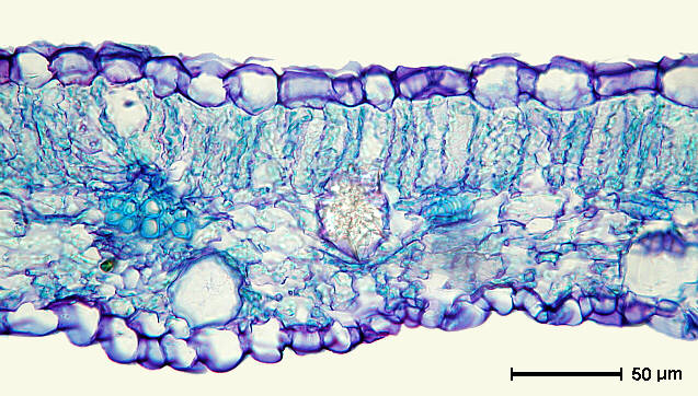
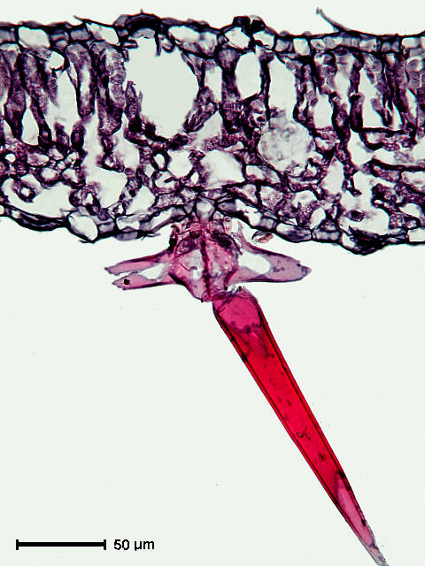
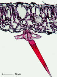 Sections by Ali G. Davalos; amphistomatic lamina and
trichome on lower (abaxial) epidermis.
Sections by Ali G. Davalos; amphistomatic lamina and
trichome on lower (abaxial) epidermis.