Basilaminar Gland - Internal Anatomy
click on the small images to open larger format images

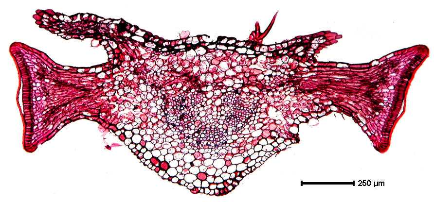
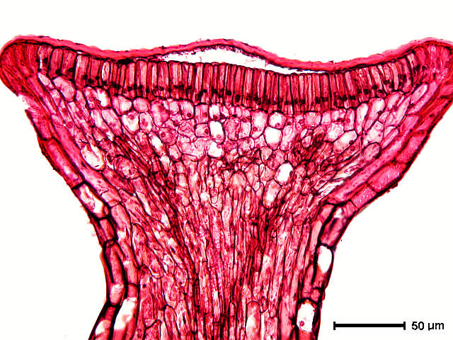
Section by Ellyn A. Dazenski. Transverse section at
junction of petiole and lamina.

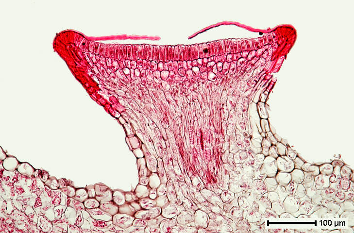
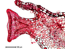 Sections by Cathy E. McLane (left) and Ellyn A. Dazenski
(right); darkly stained cells in the gland stalk represent vascular
supply to the secretory cells.
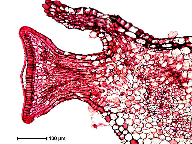
Sections by Cathy E. McLane (left) and Ellyn A. Dazenski
(right); darkly stained cells in the gland stalk represent vascular
supply to the secretory cells.
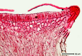
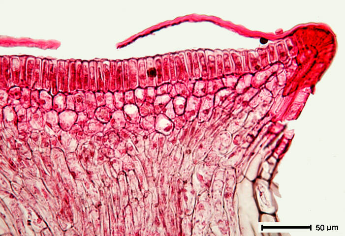
 Sections by Cathy E. McLane (left) and Ellyn A. Dazenski
(right); nectar secretion may cause the separation of cuticle
from the outer periclinal walls of the palisade-like cells forming
the secretory epidermis; it may be necessary for the cuticle to
rupture before the nectar can be gathered by insects (probably
ants).
Sections by Cathy E. McLane (left) and Ellyn A. Dazenski
(right); nectar secretion may cause the separation of cuticle
from the outer periclinal walls of the palisade-like cells forming
the secretory epidermis; it may be necessary for the cuticle to
rupture before the nectar can be gathered by insects (probably
ants).
 Section by Ellyn A. Dazenski (right)