TIMING AND 3-DIMENSIONAL ANALYSIS OF MUSCOLOSKELETAL
DEVELOPMENT
In order to create three-dimensional reconstructions of
individual cartilages, tadpoles are serial sectioned and each section is
captured by video in the computer. From these sections, individual cartilages
are isolated and traced, and binary images created from each section are
stacked to create the three dimensional reconstruction.
Sample Sections of Leptodactylus gracilis
Tadpole at Stage 32
Some individual cartilages are labeled.
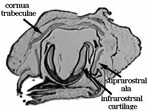
(Above)- Section through anterior portion
of chondrocranium.
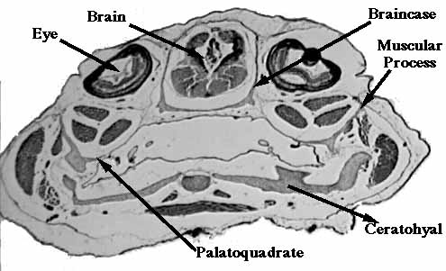
(Above)- Section through middle portion of
chondrocranium.
Last modified January 9, 1998.
Return Home