wjh home
Research Projects - W. John Hayden
Past to Present, in Approximate Chronological Order
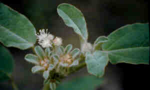
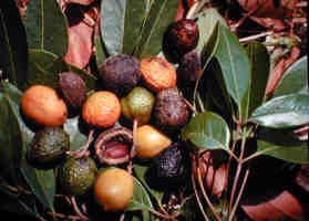 Picrodendron - This small tree, known to some as the
Jamaica Walnut occurs on coastal limestones of Cuba, Hispaniola,
Jamaica, and some of the Cayman and Bahama Islands. Its first
appearance in the scientific literature dates to the late 17th
century when Sir Hans Sloane wrote a brief account of it from
his experiences in Jamaica. Sloane suggested relationships with
Juglandaceae and various botanists over the centuries have made
their own suggestions, ranging from assignment in three different
genera and some nine different families. My master's thesis at
the University of Maryland included a detailed review of the botanical
history of Picrodendron, a thorough description of the
anatomical structure of its leaves, nodes, and wood, and a discussion
of relationships. I concluded that the anatomical and morphological
evidence confirmed contemporary palynological work, indicating
placement in Euphorbiaceae, subfamily Oldfieldioideae. I've been
studying euphorbs ever since--it was this plant that set the hook!
Picrodendron - This small tree, known to some as the
Jamaica Walnut occurs on coastal limestones of Cuba, Hispaniola,
Jamaica, and some of the Cayman and Bahama Islands. Its first
appearance in the scientific literature dates to the late 17th
century when Sir Hans Sloane wrote a brief account of it from
his experiences in Jamaica. Sloane suggested relationships with
Juglandaceae and various botanists over the centuries have made
their own suggestions, ranging from assignment in three different
genera and some nine different families. My master's thesis at
the University of Maryland included a detailed review of the botanical
history of Picrodendron, a thorough description of the
anatomical structure of its leaves, nodes, and wood, and a discussion
of relationships. I concluded that the anatomical and morphological
evidence confirmed contemporary palynological work, indicating
placement in Euphorbiaceae, subfamily Oldfieldioideae. I've been
studying euphorbs ever since--it was this plant that set the hook!
Hayden, W. J. 1977. Comparative
anatomy and systematics of Picrodendron, genus incertae sedis.
J. Arnold Arbor. 53: 257-279.
Hayden, W. J., & J. L. Reveal.
1980. Proposal for the conservation of the generic name Picrodendron
Grisebach (1859) against Picrodendron Planchon (1846) (Euphorbiaceae).
Taxon 29: 507-511.
Hayden, W. J., W. T. Gillis,
D. E. Stone, C. R. Broome, & G. L. Webster. 1984. Systematics
and palynology of Picrodendron: further evidence for relationship
with Oldfieldioideae (Euphorbiaceae). J. Arnold Arbor. 65: 105-127.
Oldfieldioideae - wood and
leaf anatomy - My doctoral
dissertation research (University of Maryland) was a natural extension
of my masters project. As noted above, my work on the enigmatic
genus Picrodendron suggested that it should be classified
in this subfamily. Since I had only examined only a few specimens
of Oldfieldioideae, and since the subfamily seemed to consist
of an odd assemblage of taxa strewn across the former continental
land mass of Gondwana, I thought that a thorough survey would
prove interesting. Results suggest that the subfamily is indeed
anatomically distinct from subfamily Phyllanthoideae, within which
most oldfieldioid genera had been classified previously. Moreover,
Oldfieldioideae was found to consist of two major clades, one
from Africa and South America consisting largely of taxa with
compound leaves, and the other, with simple leaves, centered in
Australasia. In addition, several other genera appear to form
an ill-defined basal cluster within the subfamily.
Hayden, W. J. 1992. Wood anatomy
and relationships of Australasian Oldfieldioideae (Euphorbiaceae).
Presented at symposium on "Diversity of Pacific Basin Woods
in Past, Present, and Future," sponsored by the National
Tropical Botanical Garden, Lawai, Hawaii, and the International
Association of Wood Anatomists.
Hayden, W. J. 1994. Systematic
anatomy of Oldfieldioideae (Euphorbiaceae). I. Overview. Ann.
Missouri Bot. Gard. 81: 180--202.
 Neowawraea
- While preparing various wood specimens from Phyllanthoideae
for comparison with oldfieldioid woods, I happened to section
a specimen of Drypetes phyllanthoides from the Hawaiian
Islands. This plant had been originally discovered by Joseph Rock
and named Neowawraea phyllanthoides, but it had been re-classified
as a species of Drypetes by Earl Edward Sherff. I had sectioned
numerous woods from other species of Drypetes before and
all
Neowawraea
- While preparing various wood specimens from Phyllanthoideae
for comparison with oldfieldioid woods, I happened to section
a specimen of Drypetes phyllanthoides from the Hawaiian
Islands. This plant had been originally discovered by Joseph Rock
and named Neowawraea phyllanthoides, but it had been re-classified
as a species of Drypetes by Earl Edward Sherff. I had sectioned
numerous woods from other species of Drypetes before and
all  were hard, flinty, and technically
difficult to convert into permanent microscope slides. Drypetes
(Neowawraea) phyllanthoides was totally different. It cut
like butter and was a breeze to process. Not surprisingly, its
wood structure proved to be completely different from every other
Drypetes, which is what an honors student (see citations)
helped me to do in my early years at UR. We concluded that this
plant was better better returned to the genus Neowawraea.
When I presented this study at a joint session of ASPT and BSA,
I was approached by a botanist then working on a revision of the
Hawaiian flora. It seems an amateur botanist in Hawaii had just
secured the first ever collection of pistillate flowers of Neowawraea
and he offered to let me study them, especially if I would agree
to
were hard, flinty, and technically
difficult to convert into permanent microscope slides. Drypetes
(Neowawraea) phyllanthoides was totally different. It cut
like butter and was a breeze to process. Not surprisingly, its
wood structure proved to be completely different from every other
Drypetes, which is what an honors student (see citations)
helped me to do in my early years at UR. We concluded that this
plant was better better returned to the genus Neowawraea.
When I presented this study at a joint session of ASPT and BSA,
I was approached by a botanist then working on a revision of the
Hawaiian flora. It seems an amateur botanist in Hawaii had just
secured the first ever collection of pistillate flowers of Neowawraea
and he offered to let me study them, especially if I would agree
to  write up the genus for his flora
project. I agreed on the spot, thus initiating phase two of my
adventures with Neowawraea, focused largely on herbarium
specimens and the newly obtained pistillate flowers. Sheila Hayden
provided me with the first ever technical illustration of the
plant and after taking a single glance at her drawings, a colleague
made the seminal suggestion that Neowawraea looked an awful
lot like plants in the genus Flueggea. As it turned out,
Neowawraea shared a distinctive pit in the seed coat known
elsewhere in the euphorbs only among species of Flueggea.
Clearly, Neowawraea phyllanthoides was in reality a species
of Flueggea. Since the name Flueggea phyllanthoides
had already been published for yet another plant, the Hawaiian
entity became known as Flueggea neowawraea W. J. Hayden.
Flueggea neowawraea is extremely rare and in danger of
extinction.
write up the genus for his flora
project. I agreed on the spot, thus initiating phase two of my
adventures with Neowawraea, focused largely on herbarium
specimens and the newly obtained pistillate flowers. Sheila Hayden
provided me with the first ever technical illustration of the
plant and after taking a single glance at her drawings, a colleague
made the seminal suggestion that Neowawraea looked an awful
lot like plants in the genus Flueggea. As it turned out,
Neowawraea shared a distinctive pit in the seed coat known
elsewhere in the euphorbs only among species of Flueggea.
Clearly, Neowawraea phyllanthoides was in reality a species
of Flueggea. Since the name Flueggea phyllanthoides
had already been published for yet another plant, the Hawaiian
entity became known as Flueggea neowawraea W. J. Hayden.
Flueggea neowawraea is extremely rare and in danger of
extinction.
Brandt, D. S., & W. J. Hayden. 1983. Wood anatomy and
relationships of the Hawaiian endemic genus Neowawraea
(Euphorbiaceae). Virginia Academy of Science Annual Meeting,
Fairfax, Virginia. ABSTRACT: Virginia J. Sci. 34: 138.
Hayden, W. J., & D. S. Brandt. 1984. Wood anatomy and
relationships of Neowawraea (Euphorbiaceae). A.I.B.S.
Annual Meeting, Fort Collins, Colorado. ABSTRACT: Amer. J. Bot.
71(no.5, part 2): 170.
Hayden, W. J., & D. S. Brandt. 1984. Wood anatomy and
relationships of Neowawraea (Euphorbiaceae). Syst. Bot.
9: 458-466.
Hayden, W. J. 1986. Floral morphology and relationships of
Neowawraea (Euphorbiaceae). Virginia Academy of Science
Meeting, Harrisonburg, Virginia. ABSTRACT: Virginia J. Sci. 37:
78.
Hayden, W. J. 1987. The identity of the genus Neowawraea
Rock (Euphorbiaceae). A.I.B.S. Annual Meeting, Columbus, Ohio.
ABSTRACT: Amer. J. Bot. 74: 736.
Hayden, W. J. 1987. The identity of the genus Neowawraea
(Euphorbiaceae). Brittonia 39: 268-277.
Hayden, W. J. 1990. Flueggea Willd. Pp. 620-621. In:
W. L. Wagner, S. H. Sohmer & D. Herbst, Manual of the Flowering
Plants of Hawai'i. University of Hawaii Press and Bishop Museum
Press, Honolulu.
Opportunities
for student projects with Flueggea (Neowawraea) - 1. Establishment of Flueggea neowawraea
germplasm in cultivation (will be absolutely dependent upon securing
co-operation and guidance from botanists in Hawaii. 2. Survey
of leaf anatomy in the genus Flueggea with a focus on
the interplay of ecological/environmental adaptations and relationships.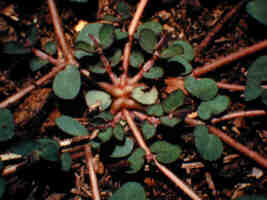
Chamaesyce - In
the course of my work with various euphorbiaceous plants I eventually
became acquainted with the genus Chamaesyce, a distinctive
group of weedy, C4 photosynthesizers postulated long ago to be
derived from species of Euphorbia via a straightforward
reduction process so that the whole plant body in Chamaesyce
could be interpreted to be homologous with the reproductive portion
(pleiochasium) of species in Euphorbia subgenus Esula
or subgenus Agaloma. With funding from the Jeffress Trust,
I undertook a comparative and developmental study of the key structures
putatively thought to be homologous, the plexus of branches that
forms at ground level in Chamaesyce and the whorl of branches
that signal the transition to the flowering phase in species of
Euphorbia. In short, the two regions are developmentally
and anatomically dissimilar and, thus, the long-held putative
homology is not supported by anatomical evidence. To date, published
materials document this conclusion for Chamaesyce maculata,
a typical prostrate species, and several species with erect growth
habits (C. hirta, C. hypericifolia, and C. 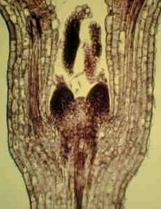 mesembrianthemifolia).
The latter study included SEM images of critical developmental
stages and included much work done by an undergraduate student
(see citations, below).
mesembrianthemifolia).
The latter study included SEM images of critical developmental
stages and included much work done by an undergraduate student
(see citations, below).
 In
the course of germinating seeds of Chamaesyce maculata
I observed the rapid formation of a mucilage layer on the seed
coat in response to water. I had never heard of a thing at the
time, and was quite chagrined when, a few months later, a paper
appeared describing the cells and structures involved in exactly
the same species formidable detail. But I had also observed that
another species, C. mesembrianthemifolia, failed to form
mucilage, even after prolonged exposure to water. I also knew
that these two species were classified in different sections of
the genus Chamaesyce, so with the help of an undergraduate
student (see citations below) we undertook a survey of mucilaginous
testa throughout the genus, and found its occurrence to be reasonably
well-correlated with the major taxonomic divisions of Chamaesyce.
Further, via SEM, we were able to correlate mucilage production
with other structural details of the testa.
In
the course of germinating seeds of Chamaesyce maculata
I observed the rapid formation of a mucilage layer on the seed
coat in response to water. I had never heard of a thing at the
time, and was quite chagrined when, a few months later, a paper
appeared describing the cells and structures involved in exactly
the same species formidable detail. But I had also observed that
another species, C. mesembrianthemifolia, failed to form
mucilage, even after prolonged exposure to water. I also knew
that these two species were classified in different sections of
the genus Chamaesyce, so with the help of an undergraduate
student (see citations below) we undertook a survey of mucilaginous
testa throughout the genus, and found its occurrence to be reasonably
well-correlated with the major taxonomic divisions of Chamaesyce.
Further, via SEM, we were able to correlate mucilage production
with other structural details of the testa.
Another student (see citations)
completed a systematic revision of a group of Chamaesyce
species known as the "Pleiadenieae" for his honors thesis.
This group was of critical interest because of their supposed
intermediate position between Euphorbia and the rest of
the genus Chamaesyce. These plants had not received comprehensive
critical study since the 1860s and 1870s.
Rosengarten, M., & W. J.
Hayden. 1983. Stem ontogeny in Chamaesyce hirta (Euphorbiaceae).
Virginia Academy of Science Annual Meeting Fairfax, Virginia.
ABSTRACT: Virginia J. Sci. 34: 142.
Hayden, W. J. 1985. Stem ontogeny
and phylogeny in Chamaesyce. Virginia Academy of Science
Meeting, Williamsburg, Virginia. ABSTRACT: Virginia J. Sci. 36:
119.
Hayden, W. J. 1988. Ontogeny
of the cotyledonary region of Chamaesyce maculata (Euphorbiaceae).
A.I.B.S. Annual Meeting, Davis, Calif. ABSTRACT: Amer. J. Bot.
75(no. 6, pt. 2): 178.
Hayden, W. J. 1988. Ontogeny
of the cotyledonary region of Chamaesyce maculata (Euphorbiaceae).
Amer. J. Bot. 75: 1701-1713.
McGuire, W. & W. J. Hayden.
1990. Anatomical comparison of woody tissues of Chamaesyce
nutans and C. hypericifolia (Euphorbiaceae). Virginia
Academy of Science Meeting, Fairfax, Virginia. ABSTRACT: Virginia
J. Sci. 41: 67.
Jordan, M. S., & W. J. Hayden.
1992. Anatomy of the seed surface in Chamaesyce: preliminary
studies of the mucilaginous layer. Virginia Academy of Science
Meeting, Richmond, Virginia. ABSTRACT: Virginia J. Sci. 43: 238.
Jordan, M. S., & W. J. Hayden.
1992. A survey of mucilaginous testa in Chamaesyce. Collect.
Bot. (Barcelona) 21: 79-89.
Simmons, M. P., & W. J. Hayden.
1994. Systematics of Chamaesyce subsection Pleiadeniae
(Euphorbiaceae). Virginia Academy of Science Meeting, James Madison
University, Harrisonburg, Virginia. ABSTRACT: Virginia J. Sci.
45: 72.
Hayden, W. J. & O. G. Troyanskaya.
1997. Seedling development in two species of Chamaesyce
with erect growth habits. Virginia Academy of Science Meeting,
Virginia Tech, Blacksburg, VA. ABSTRACT: Virginia J. Sci. 48:96.
Simmons, M. P., & W. J. Hayden.
1997. Revision of the cerrado hemicryptophyte Chamaesyce
of Boissier's "Pleiadeniae" (Euphorbiaceae). Brittonia.
49: 155--180.
Hayden, W. J. & O. Troyanskaya.
1998. Seedling development in species of Chamaesyce with
erect growth habits. Sida 18: 43--55.
Opportunities
for student projects with Chamaesyce
- Assistance is needed for studies of seedling ontogeny in species
of the "Pleiadeniae" group (native to southern Brazil
and adjacent regions), the Acutae group (native to Texas and
adjacent Mexico), and certain Hawaiian species; acquisition of
suitable specimens is a pre-requisite for making progress with
any of these potential projects.
Floristic inventories - Although the plants of Virginia are
reasonably well known, in general, our knowledge of the geographic
distribution of plants (exactly which plants occur in what parts
of the state) is still somewhat sketchy. This knowledge is obtained
by the collection and curation of herbarium specimens, the raw
data of systematics and biogeography. Some parts of the state
have been well-collected while others are much less well known.
Further, comprehensive collecting projects are excellent means
for students to become knowledgeable about local plant diversity,
and the collections produced, when added to an herbarium, become
useful materials for teaching and research. Below are citations
related to two floristic inventories that I and my students have
pursued to closure.
Hayden, W. J., & M. F. Johnson.
1986. Flora of Richmond National Battlefield Park (Chickahominy
Bluffs, Cold Harbor, Fort Darling, Fort Harrison, Garthright
House, and Watt House units). U.S. Department of the Interior,
National Park Service, Research/Resources Management Report MAR-16,
40 pp.
Hayden, W. J., M. L. Haskins,
& M. F. Johnson. 1988. Flora of Richmond National Battlefield
Park (Beaver Dam Creek, Malvern Hill, and Parker's Battery units).
U.S. Department of the Interior, National Park Service, Research/Resources
Management Report, MAR-27, 33 pp.
Hayden, W. J., M. L. Haskins,
M. F. Johnson, & J. M. Gardner. 1988. Flora of Richmond National
Battlefield Park, Virginia. Virginia Academy of Science Meeting,
Charlottesville, Virginia. ABSTRACT: Virginia J. Sci. 39: 126.
Hayden, W. J., M. L. Haskins,
M. F. Johnson, & J. M. Gardner. 1989. Flora of Richmond National
Battlefield Park, Virginia. Castanea 54: 87-104.
Simmons, M. P., D. M. E. Ware,
& W. J. Hayden. 1993. The vascular flora of the Potomac River
drainage of King George County, Virginia. Virginia Academy of
Science Meeting, Old Dominion University, Norfolk, Virginia.
ABSTRACT: Virginia J. Sci. 44: 123.
Simmons, M. P., D. M. E. Ware,
& W. J. Hayden. 1995. The vascular flora of the Potomac River
watershed of King George County, Virginia. Castanea 60: 179--209.
Opportunities
for student projects with floristic inventories
- Almost unlimited; any reasonable sized area with natural vegetation
may prove of interest.
Penthorum - My
first graduate student (see citations, below) wanted to work on
a wetland plant from Virginia and Penthorum seemed to fit
the bill. Penthorum sedoides--the "Ditch Stonecrop"--occurs
along the margins of bodies of water throughout eastern North
America. Another species, Penthorum chinensis, occurs in
similar habitats in eastern Asia. The systematic placement of
Penthorum was in doubt and there had been no thorough study
of its anatomical structure, so my student's goal was to provide
a thorough description of wood, node, and leaf of both species
and assess what the anatomical evidence indicated in terms of
relationships with Saxifragaceae and Crassulaceae, the two families
thought to be most closely related. In general, she found that
Penthorum lacked the distinctive synapomorphous features
that might be used to define either Saxifragaceae or Crassulaceae.
Thus, no compelling argument could be made to place it in either
family, and we argued that the monotypic family Penthoraceae should
be resurrected to accommodate this genus.
The gynoecium of Penthorum
always seemed peculiar to me. Over a period of years in the 90's,
I used Penthorum as the basis for several class projects,
which led, eventually, to work by some additional undergraduate
students on the anatomy and morphology of the gynmoecium of Penthorum.
Contrary to the usual phyllosporous condition long-theorized to
be the ancestral condition for flowering plants as a whole, we
interpret the gynoecium of Penthorum to be stem-borne,
or stachysporous. Moreover, preliminary studies reveal long-overlooked
evidence for variability in carpel number in Penthroum sedoides.
Haskins, M. L., & W. J. Hayden.
1985. Anatomy and systematics of the ditch stonecrop, Penthorum.
Virginia Academy of Science Meeting, Williamsburg, Virginia.
ABSTRACT: Virginia J. Sci. 36: 119.
Haskins, M. L., & W. J. Hayden.
1985. Anatomy and systematics of Penthorum. A.I.B.S. Annual
Meeting, Gainesville, Florida. ABSTRACT: Amer. J. Bot. 72(no.
6, pt. 2): 956.
Haskins, M. L., & W. J. Hayden.
1987. Anatomy and affinities of Penthorum. Amer. J. Bot.
74: 162-175.
Hayden, W. J. & J. D. Lewandowski.
1997. Gynoecium structure in Penthorum. Virginia Academy
of Science Meeting, Virginia Tech, Blacksburg, VA. ABSTRACT:
Virginia J. Sci. 48: 95.
Hayden, W. J. & J. D. Lewandowski.
1997. Anatomy and morphology of the gynoecium of Penthorum.
A.I.B.S. Annual Meeting, Montreal, Canada. ABSTRACT: Amer. J.
Bot. 84(6, Supplement): 201.
Opportunities
for student projects with Penthorum
- Assistance is needed in documentation of asynchrony in ovule
development, and variability of carpel number.
Methyl salicylate roots of
violets - My plant systematics
class includes what I call "key-out guizzes," unknowns
that the students must identify using the keys found in standard
floristic works. During one spring semester in the late 1980s
I happened to use a particular species of stemmed violet as an
unknown. Most students were able to proceed accurately through
the key to the violets, but, unexpectedly, many had trouble with
the last few steps leading to the exact species at hand. It seems
that the roots of the unknown specimen had a remarkably strong
odor of oil of wintergreen, but this fact was indicated in the
key only for a close relative. Many students reached the wrong
identification because of this misleading shortcoming in the book.
But, as the saying goes, there are no problems, only opportunities.
As the class and I sorted through the confusion, it seemed likely
that several closely related violets in subgenus Melanium
all have wintergreen-like contents in their roots. One particularly
enthusiastic student from that class (see citations, below) decided
to straighten things out, at least for the species readily available
in Virginia. With help from a colleague in the Department of Chemistry,
we determined that the compound was indeed oil of wintergreen
(methyl salicylate), that it is found in both Viola arvensis
and V. rafinesquii, and that the compound occurs in
specialized secretory cells located in the endodermis layer of
the root.
Hayden, W. J., & J. Clough.
1989. Identification and localization of methyl salicylate in
roots of Viola subgenus Melanium. Virginia Academy of
Science Meeting, Richmond, Virginia. ABSTRACT: Virginia J. Sci.
40: 63.
Hayden, W. J., & J. Clough.
1990. Methyl salicylate secretory cells in roots of Viola
arvensis and V. rafinesquii (Violaceae). Castanea
55: 65-70.
Amanoa - While sectioning leaves of various
phyllanthoids in graduate school, I happened across the remarkable
sclereid-containing epidermal cells of Amanoa. The student
who initially worked on Neowawraea (above) also initiated
 an anatomical survey of the leaves of Amanoa,
working with leaf fragments generously provided by the Smithsonian
Institution. From what we know now, it appears that each species
in the genus has its own distinct structural pattern of sclerified
cells. But initial results from the student's anatomical studies
were murkey and we gradually realized that the problem stemmed
from a number of gross mis-identification of specimens in the
collections of the Smithsonian Institution.
an anatomical survey of the leaves of Amanoa,
working with leaf fragments generously provided by the Smithsonian
Institution. From what we know now, it appears that each species
in the genus has its own distinct structural pattern of sclerified
cells. But initial results from the student's anatomical studies
were murkey and we gradually realized that the problem stemmed
from a number of gross mis-identification of specimens in the
collections of the Smithsonian Institution.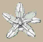 At which point, I resolved to undertake a thorough study of the
genus, encompassing all available collections from North American,
South American, and European herbaria. My work has progressed
to the point recognizing and naming several new species, an account
of the genus for the "Flora of the Venezuelan Guyana,"
a published study of wood anatomy in the genus, a draft of a detailed
monograph of the genus and drafts of floristic treatments of the
genus for Colombia and the Guianas. The original sclereid study
remains unpublished and awaits the inclusion of SEM images.
At which point, I resolved to undertake a thorough study of the
genus, encompassing all available collections from North American,
South American, and European herbaria. My work has progressed
to the point recognizing and naming several new species, an account
of the genus for the "Flora of the Venezuelan Guyana,"
a published study of wood anatomy in the genus, a draft of a detailed
monograph of the genus and drafts of floristic treatments of the
genus for Colombia and the Guianas. The original sclereid study
remains unpublished and awaits the inclusion of SEM images.
Hayden, W. J. 1990. Notes on
neotropical Amanoa (Euphorbiaceae). Brittonia 42: 260-270.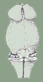
Hayden, W. J. 1991. Taxonomic
studies of Amanoa (Euphorbiaceae). Virginia Academy of
Science Meeting, Blacksburg, Virginia. ABSTRACT: Virginia J.
Sci. 42: 183.
Hayden, W. J., M. P. Simmons,
& L. Swanson. 1993. Wood anatomy of Amanoa (Euphorbiaceae).
IAWA Jour. 14: 1-9.
Hayden, W. J. 1999. Amanoa
(Euphorbiaceae). Pp. 95–99, in: J. A. Steyermark et al.,
eds., Flora of the Venezuelan Guayana, Missouri Botanical Garden
Press.
Opportunities
for student projects with Amanoa
- Assistance is needed with SEM studies of leaf features, producing
publishable distribution maps for all species, and cladistic
analyses of species within the genus.
Tetracoccus - Tetracoccus is a small genus of
desert shrubs found in southern California, Arizona, and Mexico.
It is the only member of Euphorbiaceae subfamily Oldfieldioideae
found in the US. Thus, I was pleased to volunteer to produce a
treatment for Tetracoccus for the Flora North America project
and for the Vascular Plants of Arizona project.
Hayden, W. J. 1996. The genus Tetracoccus in North
America. Virginia Academy of Science Meeting, Virginia Commonwealth
University, Richmond, Virginia. ABSTRACT: Virginia J. Sci. 47:
102.
Hayden, W. J. (in press). Tetracoccus. In: L. Landrum,
C. Mason, D. Pinkava, J. Reeder, and R. Van Devender, eds., Vascular
Plants of Arizona.
Hayden, W. J. (in press). Tetracoccus Parry. In: Flora
North America Editorial Committee (eds), Flora of North America.
Oxford University Press.

 Croton -
Stems of Croton, one of the largest and most diverse genera
in Euphorbiaceae, are characterized by several unusual anatomical
features derived from bicollateral bundles with the potential
to form two distinct cambia, the innermost of which can result
in the formation of medullary bundles. Aside from a single survey
published over a century ago, and work from our lab, virtually
no other reports of these unusual features exist.
Croton -
Stems of Croton, one of the largest and most diverse genera
in Euphorbiaceae, are characterized by several unusual anatomical
features derived from bicollateral bundles with the potential
to form two distinct cambia, the innermost of which can result
in the formation of medullary bundles. Aside from a single survey
published over a century ago, and work from our lab, virtually
no other reports of these unusual features exist. 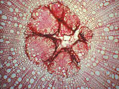
Hayden, S. M., & W. J. Hayden.
1993. Stem anatomy and medullary bundles of Croton glandulosus
var. septentrionalis (Euphorbiaceae). Virginia Academy
of Science Meeting, Old Dominion University, Norfolk, Virginia.
ABSTRACT: Virginia J. Sci. 44: 119.
Hayden, S. M., & W. J. Hayden.
1994. Stem development, medullary bundles, and wood anatomy of
Croton glandulosus var. septentrionalis (Euphorbiaceae).
IAWA Jour. 15: 51-63.
Discocarpus - Interest in this small genus from Amazonia
stemmed from the fact that it and Amanoa appear to be the
only genera in Euphorbiaceae subfamily Phyllanthoideae with sclereids
in foliar epidermis. Graduate student Sheila Hayden did a revision
of the genus, including naming one new species. She also did a
comparative study of leaf anatomy for the three species she recognized
in the genus.
Hayden, S. M., & W. J. Hayden.
1996. A revision of Discocarpus (Euphorbiaceae). Ann.
Missouri Bot. Gard. 83: 153--167.
Saururus - Saururus cernuus is a wetland
plant found throughout eastern North America. It is known as Lizard's
Tail, for its distinctive recurved inflorescence. Whereas all
previous literature had suggested that rhizomes and stems of Saururus
possess only primary growth, a recently published study of
the anatomy of Saururaceae interpreted the bundles to contain
secondary xylem. To help resolve the apparent discrepancy between
the older and more recent literature, an undergraduate student
(see citation, below) undertook a developmental study to document
the progressive accumulation of xylem elements from rhizome tips
to mature regions several years old. Whereas locally collected
Virginia material show scant evidence of secondary xylem, a sample
from Louisiana was reasonably consistent with Carlquist's report.
Hayden, W. J. & L. S. Machut.
1997. Does the rhizome of Saururus cernuus undergo secondary
growth? Virginia Academy of Science Meeting, Virginia Tech, Blacksburg,
VA. ABSTRACT: Virginia J. Sci. 48: 96.
Machut, L. S. & W. J. Hayden.
1998. Further studies of xylem development in the rhizomes of
Saururus cernuus. Virginia Academy of Science Meeting,
George Mason University, Fairfax, VA. ABSTRACT: Virginia J. Sci.
49: 70.
Opportunities
for student projects with Saururus
- More studies focusing on geographic and edaphic influences
on secondary xylem formation in the rhizomes are needed.
Chonocentrum - While working on Discocarpus,
attention was focused on this poorly known genus from the upper
Rio Negro region of Brazil. To the best of our knowledge, Chonocentrum
was collected only once, in the 1850s. There are no published
illustrations of this plant, and we believe that it has been mis-placed
taxonomically.
Hayden, W. J., & S. M. Hayden. 1996. Two enigmatic biovulate
Euphorbiaceae from the neotropics: relationships of Chonocentrum
and the identity of Phyllanoa. A.I.B.S. Annual Meeting,
Seattle, Washington. ABSTRACT: Amer. J. Bot. 83(6-supplement):
162.
Phyllanoa - Studies of Chonocentrum brought
to light problems with Phyllanoa. This genus from moderately
high altitude in the Colombian Andes is also known from a single
collection. It has been referred to Euphorbiaceae tribe Antidesmeae
(subfamily Phyllanthoideae), however, we feel that the plant really
belongs in Violaceae.
Hayden, W. J., & S. M. Hayden.
1996. Two enigmatic biovulate Euphorbiaceae from the neotropics:
relationships of Chonocentrum and the identity of Phyllanoa.
A.I.B.S. Annual Meeting, Seattle, Washington. ABSTRACT: Amer.
J. Bot. 83(6-supplement): 162.
Hayden, W. J., & S. M. Hayden.
2000. Relationships of Chonocentrum (Euphorbiaceae). Virginia
Academy of Science Meeting, Radford University, Radford, VA.
ABSTRACT: Virginia J. Sci. 51: 95.
Acalyphoideae - wood anatomy
- At 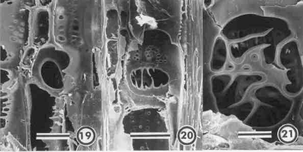 a
conference on Euphorbiaceae held at the Missouri Botanical Garden
in the early 1990s, subfamily Acalyphoideae was referred to repeatedly
as "the heart of darkness of Euphorbiaceae," a reference
to the need for comparative data to help sort out relationships
among this large and poorly known group of plants. So when I received
an invitation to present a paper at a session on wood anatomy
at the 1999 International Botanical Congress, Acalyphoideae seemed
like it would prove a worthy topic. Despite the discovery of several
genera with anatomically distinct wood structure (laticifers in
Dicoelia and one species of Dalechampia, thick-walled
fibers and scalariform perforations in tribe Galeariae, etc.)
most acalyphoid woods were too uniform to reveal systematically
useful characters. The black and white SEM images (19, 20, and
21) depict perforated ray cells with various forms of perforation
plates.
a
conference on Euphorbiaceae held at the Missouri Botanical Garden
in the early 1990s, subfamily Acalyphoideae was referred to repeatedly
as "the heart of darkness of Euphorbiaceae," a reference
to the need for comparative data to help sort out relationships
among this large and poorly known group of plants. So when I received
an invitation to present a paper at a session on wood anatomy
at the 1999 International Botanical Congress, Acalyphoideae seemed
like it would prove a worthy topic. Despite the discovery of several
genera with anatomically distinct wood structure (laticifers in
Dicoelia and one species of Dalechampia, thick-walled
fibers and scalariform perforations in tribe Galeariae, etc.)
most acalyphoid woods were too uniform to reveal systematically
useful characters. The black and white SEM images (19, 20, and
21) depict perforated ray cells with various forms of perforation
plates.
Hayden, W. J., & S. M. Hayden. 1999. Wood anatomy of Acalyphoideae
(Euphorbiaceae). Paper presented at XVI International Botanical
Congress. ABSTRACT: IAWA Journal 20: 108-109.
Hayden, W. J., & S. M. Hayden. 2000. Wood anatomy of Acalyphoideae
(Euphorbiaceae). IAWA Journal 21: 213–235.
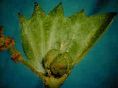
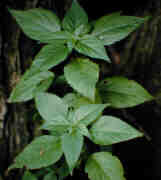
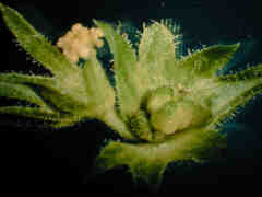
Acalypha deamii - This little plant (above, center) was
thought to be restricted to flood plain habitats within the Ohio
River drainage basin. However, quite by accident, I discovered
a population at the Huguenot Woods area of James River Park, just
a stone's throw from UR. Several seasons of field work have now
shown Acalypha deamii to be present on the James, Rappahanock,
Shenandoah, and Potomac Rivers. Reproductive structures for related
species of Acalypha are also shown above, left, A. gracilens,
right, A. rhomboidea.
Hayden, W. J. & S. M. Hayden. 1998. Anatomy of secondary
xylem in the Acalypha virginica complex. Virginia Academy
of Science Meeting, George Mason University, Fairfax, VA. ABSTRACT:
Virginia J. Sci. 49: 70.
Opportunities
for student projects with Acalypha
- Developmental studies of "normal" reproductive structures
in the Acalypha virginica complex, for comparison with
the allomorphic pistillate flowers and fruits of Acalypha
deamii.
Floristics of Yucatan, Mexico - This work has had two
focal points. First, since 2000, I have worked to document plant
diversity of the forest at Kaxil Kiuic (The Helen Moyers BioCultural
Reserve), located in the southern Puuc Region of the state of
Yucatan, Mexico. As of summer 2006, approximately 450 species.
have been documented via herbarium specimens (deposited at URV,
UADY, and CICY) and digital photographs which can be viewed at
my Flora
of Kaxil Kiuic web site. Also I am pursuing the floristics
of various members of the plant family Euphorbiaceae as they occur
throughout the Yucatan Peninsula.
wjh home